R.O.P. foundations · A practitioner’s guide
From the foot to visceral function: understanding the five bridges
How could a technique applied to the foot influence a function in the pelvis or abdomen? To examine this hypothesis, we can follow the nerve signal from the skin to the networks involved in organ control. Five “bridges” illuminate this route: five complementary arguments, with different strengths and implications.
The pathway to keep in mind
Skin of the foot
Pressure deforms the skin and can activate mechanoreceptors: sensors that respond to mechanical forces.
Sensory nerves
They carry signals towards the central nervous system. This incoming information is called afferent input.
Spinal cord and brain
Their networks process incoming information. They can change the commands sent to organs.
A bodily response
An outgoing, or efferent, signal can modify muscle or visceral activity. The response depends on the circuit involved.
Three questions to ask when reading the evidence
- Is the pathway plausible?
- Anatomy identifies pathways and connections consistent with the hypothesis.
- Has a response been measured?
- An experiment demonstrates a change with a specified stimulus under specified conditions.
- Does the patient benefit?
- A clinical study assesses a useful improvement attributable to the technique being studied.
The labels within the slides are currently in French. The guide text is available in English.
Bridge 1 / 5
Some spinal levels overlap
Can signals from the foot and pelvis reach neighbouring regions of the spinal cord?
The anatomical reference point
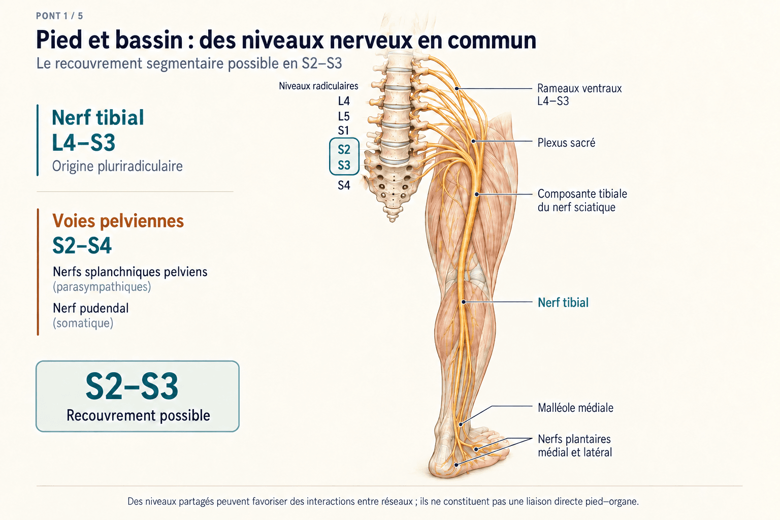
The spinal cord is organised into segments, labelled with letters including L for lumbar and S for sacral. The tibial nerve passes behind the ankle and supplies the sole. It is classically described as containing fibres from L4 to S3. The pelvic parasympathetic nerves and pudendal nerve are associated with S2–S4. There is therefore a possible overlap at S2–S3.
What anatomy describes
A peripheral nerve contains fibres from several roots. A dermatome, by contrast, is a skin territory associated mainly with one root. These maps do not coincide: plantar sensation is largely associated with S1, even though the tibial nerve as a whole includes other contributions.
What this supports
This anatomical proximity provides a coherent reason to investigate interactions between information from the foot and pelvic networks.
A limitation to keep in mind
The label “L4–S3” on a diagram does not tell us which fibres pressure on a specific area of the foot actually activates. Anatomical overlap is a starting point that needs functional observations to support it.
Bridge 2 / 5
Plantar branches can produce different effects
Could the choice of region matter, even within the territory of the tibial nerve?
The anatomical reference point
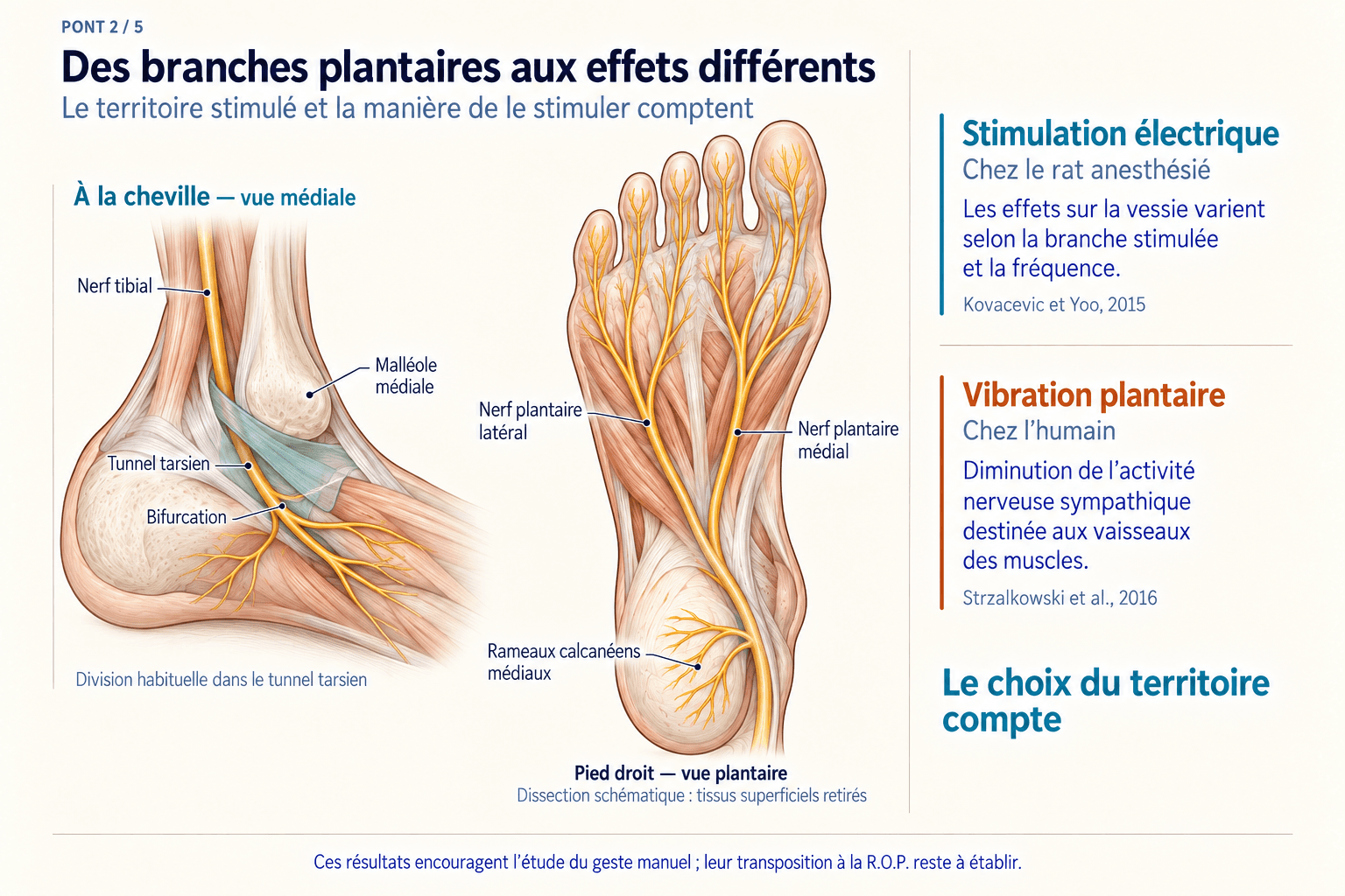
In the foot, the tibial nerve divides into branches including the medial and lateral plantar nerves, with calcaneal branches serving the heel. Here, “medial” means the big-toe side and “lateral” the little-toe side. These branches supply distinct territories, with anatomical boundaries and variations.
Electrical stimulation in rats
Kovacevic and Yoo stimulated the medial and lateral plantar nerves separately in anaesthetised rats. Bladder responses depended on the branch and frequency. Inhibition occurred at 5 and 10 Hz for the medial branch and 10 and 20 Hz for the lateral branch; at 50 Hz, the lateral branch could instead elicit an excitatory response. Hertz (Hz) means electrical pulses per second.
Mechanical stimulation in humans
Strzalkowski and colleagues showed that vibration of the sole could reduce the frequency of sympathetic nerve bursts directed to muscle blood vessels. The sympathetic system helps regulate automatic bodily functions. The measurement concerned vascular nerve activity, not the bladder. Hand vibration also produced an effect.
What this supports
The first study shows that two branches of one nerve can produce different responses. The second shows that mechanical stimulation of skin can modify autonomic nerve activity in humans. Together, they justify investigating both the territory and the way it is stimulated.
A limitation to keep in mind
These experiments answer different questions. They validate neither a point-to-organ correspondence nor the effectiveness of manual pressure. Electrical frequencies do not provide a massage rhythm to reproduce.
Bridge 3 / 5
Several nerves can provide access to the networks
Is the tibial nerve the only pathway capable of influencing a pelvic function?
The anatomical reference point
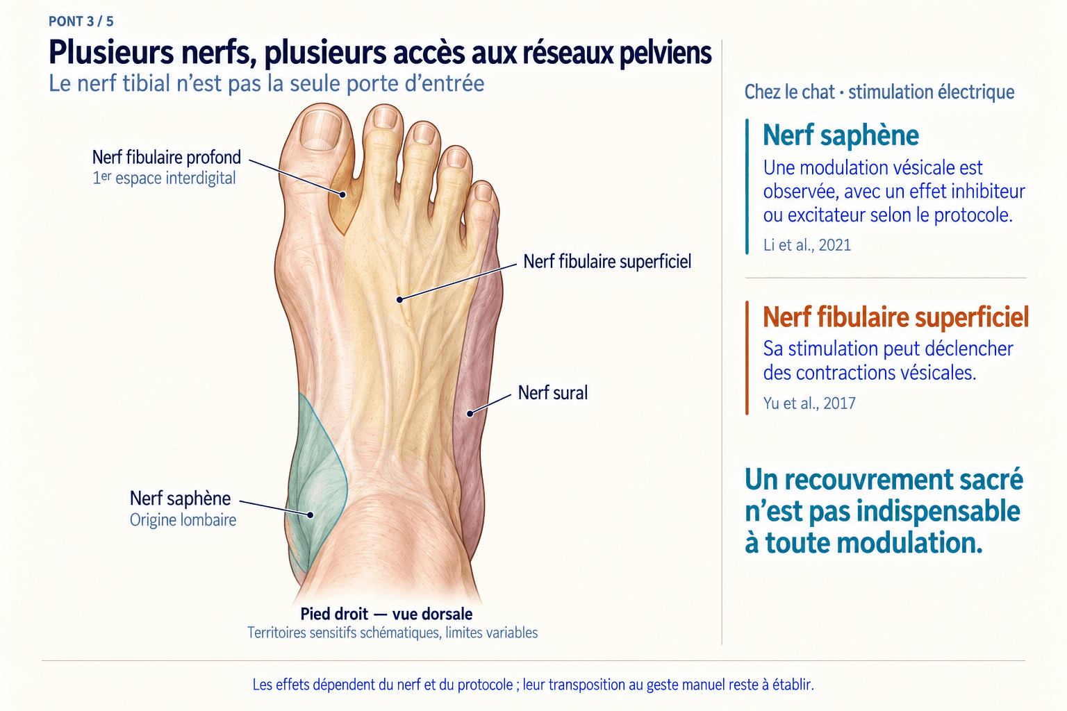
The foot also receives branches of the sural, fibular and saphenous nerves. They cover different areas and do not have identical spinal origins. The saphenous nerve arises mainly from lumbar levels, so it does not directly share the S2–S4 sacral origins described above.
Effects measured in cats
Electrical stimulation of the saphenous nerve can modify the bladder reflex, with effects depending on factors including frequency and experimental conditions. Another study shows that superficial fibular nerve stimulation can elicit bladder contractions. A sensory input can therefore have either an inhibitory or an excitatory influence.
Li et al., 2021 · saphenous nerveYu et al., 2017 · superficial fibular nerve
What this supports
Several peripheral pathways can reach circuits controlling the same function. This gives a rationale for comparing different foot territories.
A limitation to keep in mind
Root proximity alone cannot rank nerves by effectiveness. A result established for one nerve and protocol does not automatically transfer to other territories or a manual technique.
Bridge 4 / 5
The spinal cord can integrate different inputs
Where can sensory signals interact with networks related to organs?
The anatomical reference point
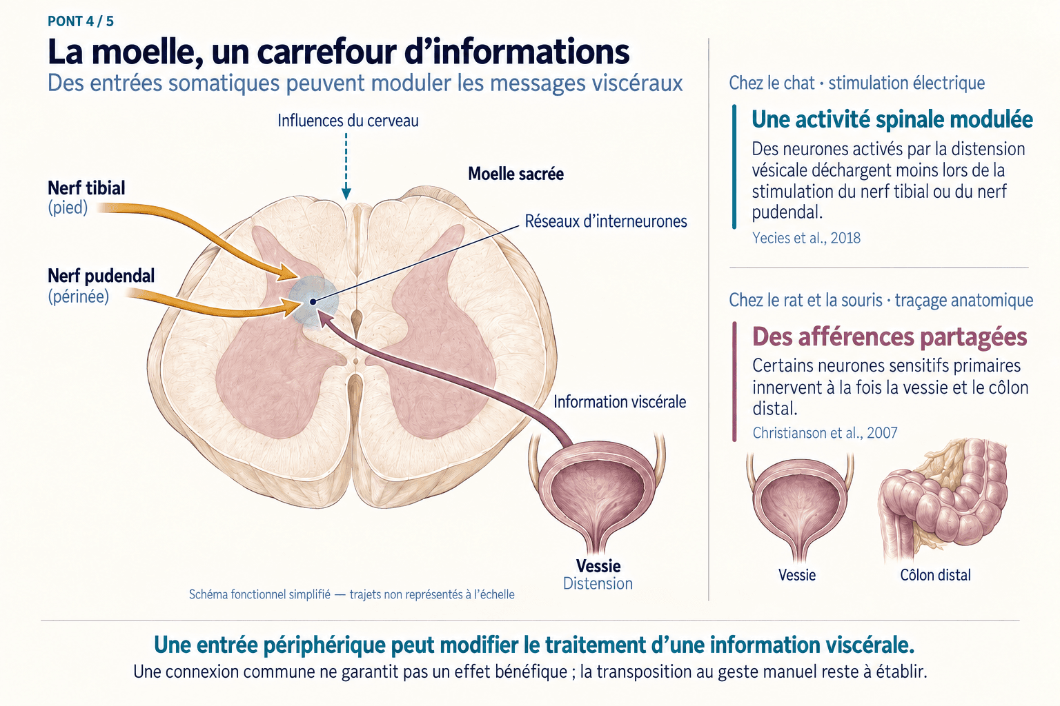
The spinal cord contains interneurons: cells that connect other neurons. Some receive or integrate several types of information. Convergence means that different inputs reach the same neuron or network. Here, “somatic” refers to skin, muscles and joints, while “visceral” refers to internal organs.
Recordings from sacral neurons in cats
Yecies and colleagues recorded the activity of spinal neurons activated by bladder distension. Electrical stimulation of either the tibial or pudendal nerve could reduce the activity of these same neurons. This is a functional observation at this level, beyond proximity on an anatomical diagram.
Connections between pelvic organs
In rats and mice, anatomical tracing also shows that some primary sensory neurons innervate both the bladder and distal colon. This shared supply helps explain interactions between organs, including increased sensitivity in one when the other is irritated.
What this supports
Pelvic networks contain sites of integration where a peripheral input can modify the processing of visceral information.
A limitation to keep in mind
An effect recorded in the spinal cord may also depend on commands from the brain. A shared connection does not necessarily imply a beneficial effect: it may contribute to inhibition, excitation or sensitisation.
Bridge 5 / 5
The brain also contributes to the response
How could an influence extend beyond neighbouring spinal levels?
The anatomical reference point
“Supraspinal” means above the spinal cord. The brainstem, between the spinal cord and the higher brain regions, helps control urination and other automatic functions. Information travels up to it and commands travel down. The autonomic nervous system includes sympathetic and parasympathetic pathways.
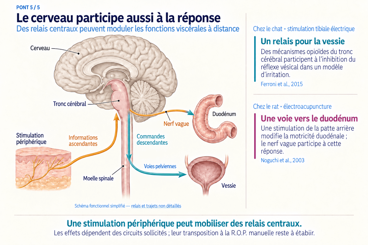
Relays investigated in animals
In cats, a tibial stimulation experiment showed that brainstem opioid mechanisms participate in inhibiting the bladder reflex in a bladder irritation model. In rats, electrical acupuncture stimulation of the hind paw modified duodenal motility; the effect depended in part on the vagus nerve, a major parasympathetic pathway to the viscera.
Ferroni et al., 2015 · bladderNoguchi et al., 2003 · duodenum
What this supports
Peripheral stimulation can engage central relays and influence a distant function. These relays may also contribute to pelvic effects.
A limitation to keep in mind
Passing through the brain does not necessarily mean a diffuse effect on all organs: circuits can remain selective. Their involvement must be established for the stimulus and function being studied; it does not by itself demonstrate an R.O.P. effect on a given organ.
What about pathways to the head?
The full article examines a sixth bridge: cranial outputs from the brainstem. It extends the fifth bridge, with hypotheses that need to be examined function by function.
The example of the pupil
In anaesthetised rats, electrical hind-paw stimulation elicited pupil dilation consistent with a reduction in the parasympathetic command that normally constricts the pupil. Dilation therefore does not necessarily mean sympathetic activation.
This experiment does not establish effects of manual techniques on glands, the pharynx or the larynx. For these structures, sharing a brainstem region is not enough to prove interaction. Parasympathetic control of glands must also be distinguished from motor control of striated muscles.
Full article · Explore bridge 6 →What the practitioner can take from this
These bridges provide converging reasons to study the influence of sensory inputs from the foot on visceral functions. The bladder is a particularly well-documented starting point in the research presented here: peripheral branches, sacral neurons, brain relays and clinical neuromodulation trials.
A clinical reference point
In the randomised SUmiT trial, 220 adults with overactive bladder received either percutaneous tibial nerve stimulation or a sham procedure. Results at 12 weeks favoured the technique being tested. It used electrical stimulation through a needle near the ankle; the trial did not assess manual R.O.P.
Clinical experience can help identify comparisons worth making. To support the practice, the next steps are to describe the territory and technique, choose a measurable function and compare results under conditions that allow an effect to be attributed to stimulation.